Cardiovascular diseases have become the number one killer to threatening human health, and coronary microcirculation disorders is an important pathogenesis and an important factor affecting the prognosis of cardiovascular diseases such as coronary heart disease and heart attack.
At present, the four major imaging techniques of ultrasound, MRI, X-ray and nuclear medicine commonly used in clinical practice can only indirectly assess microcirculatory function, but cannot visually display microvascular structures; although CT and MR devices for basic experimental small animals can already display some microvascular structures, while they cannot provide real-time dynamic imaging and cannot provide hemodynamic information.
The development of imaging devices that can visually display the structure of myocardial microvascular structure and evaluate its function in real time is a common need for clinical and basic research.
Based on this, Professor Mingxing Xie's team from the Department of Ultrasound Imaging, Union Hospital of Tongji Medical College, Huazhong University of Science and Technology, in collaboration with Professor Benpeng Zhu's team from the School of Optics and Electronic Information, Huazhong University of Science and Technology, developed a super-harmonic transducer based on PMN-PT 1-3 composite piezoelectric material and stacked structure, and developed a laser Doppler blood flow detection probe using optical beat technology, and integrated the two through micromachining technology to create a high-resolution acoustic/optical dual-mode transducer.
High-performance FPGA signal processing circuit and high-precision semi-automatic motion system are applied to acquire and process acoustic/optical signals.
The interactive dual-mode image real-time processing software are developed to realize 3D display of myocardial microvascular and blood flow.
This project innovatively integrated dual-frequency super-harmonic ultrasonography imaging technology and laser Doppler technology to develop a small animal acoustic/optical dual-mode imaging device, which is able to display 3D microscopic imaging of the myocardial microcirculation in vivo and realize real-time detection of myocardial low-velocity blood flow within a certain depth range, and provided a new tool for myocardial microcirculation imaging in vivo and hemodynamic studies.
This project was successfully funded by the National Natural Science Foundation of China for the development of major research instruments (81727805, RMB 8.9 million), which fully reflecting the advantages of cardiovascular research in Union Hospital and the technical strength of science and technology in Huazhong University of Science and Technology, it is a model of the combination among the strong ones of medical-industrial-science in our university.
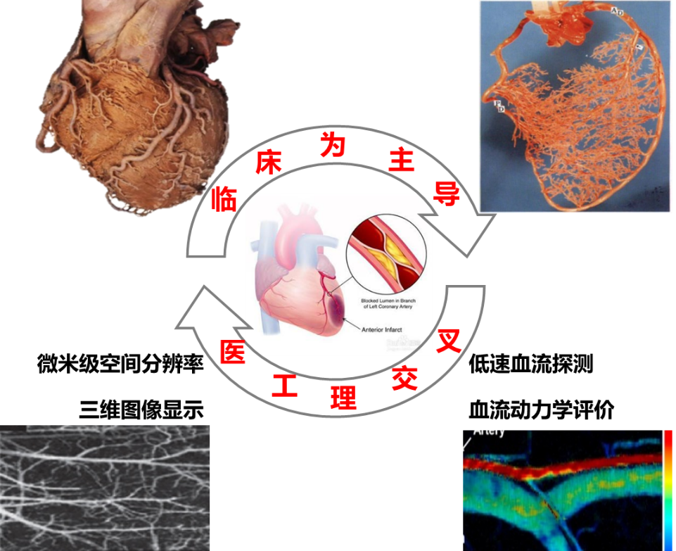
临床为主导 Clinically oriented
医工理交叉 Interdisciplinary of Medical Engineering and Science
微米级空间分辨率 Micron spatial resolution
三维图像显示 3D image display
低速血流探测 Low velocity blood flow detection
血流动力学评价 Hemodynamic evaluation


 Chinese